Jason Dowling
Team Leader
Medical Image Analysis
Our team partners with clinicians to develop and validate novel technology to, extract quantitative information from medical image data and to understand and improve disease diagnosis, treatment planning and treatment delivery.
In collaboration with clinical partners our team produce high impact scientific research in a range of medical disciplines, including oncology, cardiology, radiology, sports medicine, respiratory physiology and orthopaedic surgery. The team currently receive a number of competitive grants (including three current NHMRC MRFF grants) and industry collaboration.
Impact on the Health System
Our projects develop cutting edge validated methods to acquire and automatically extract information from medical images to improve diagnosis, treatment delivery efficiency, cost-effectiveness and patient quality of life.
Our Solutions
Our solutions have been developed in partnership with healthcare practitioners at many sites across Australia.
Case Studies
August 2, 2018Medical Imaging, Medical Imaging Case Study, SoftwareAEHRC has developed a flexible surgical simulation software platform, MILXSim, which provides high performance rendering and simulation capabilities to the Surgical Simulation and Planning team. The platform is developed in C++ and...
November 23, 2017Biomedical, Case study, Informatics, Lee Reid, Machine Learning, Medical Imaging, Medical Imaging Case Study, Neuroimaging, Research, ScienceCONSULT is a 3D experience that allows neurosurgeons to see the invisible, making brain surgery safer and more effective than ever before Brain Surgery Remains A High-Risk Procedure Brain surgery is dangerous. The...
November 17, 2017Case study, Medical Imaging Case StudyMusculoskeletal Segmentation In 2001, 6.1 million Australians (32%) had long-term arthritis or a musculoskeletal disorder with almost 1.2 million reporting an associated disability (Bhatia 2005). Osteoarthritis (OA) is the most common...
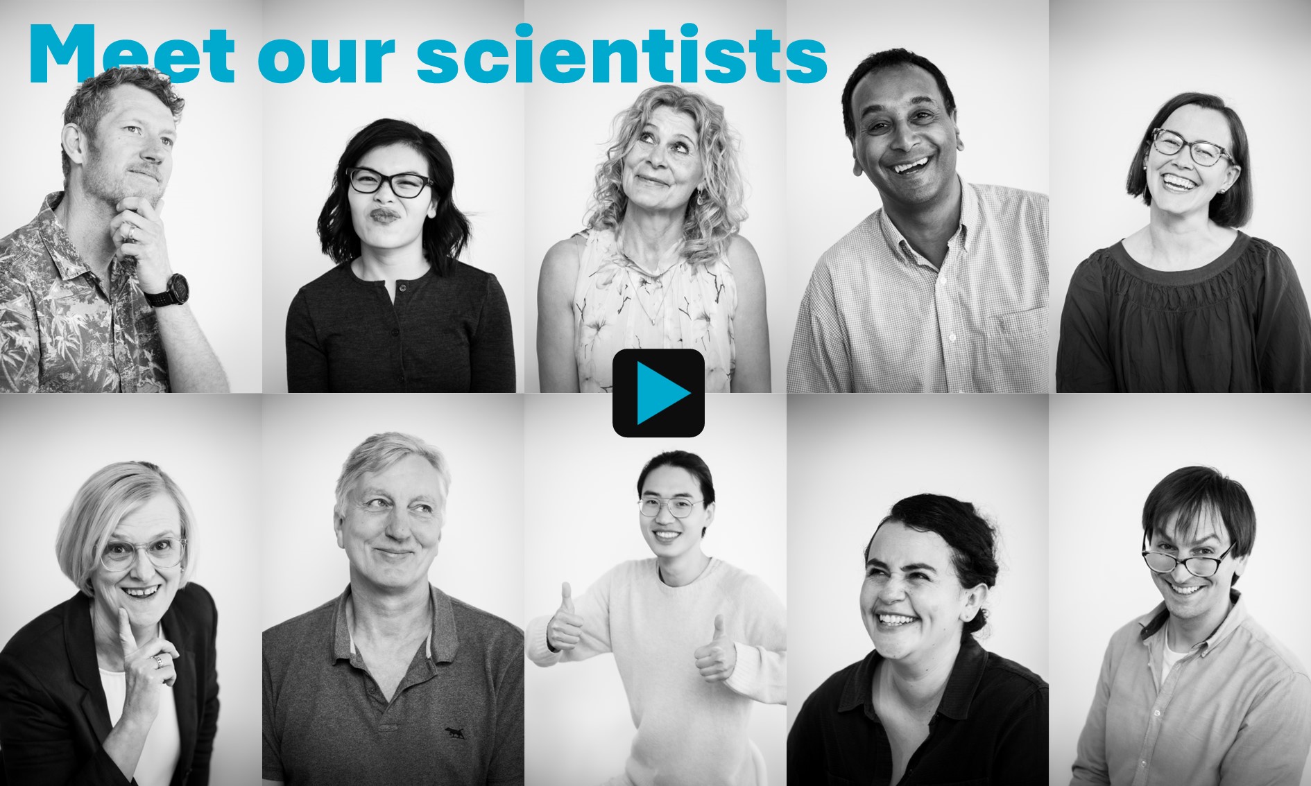
Team Members:

Jason Dowling

Ashley Gillman

Simon Puttick

Celine Leung

Jess Bugeja

Hollie Min

Rodrigo Fernandez Santa Cruz

Leo Lebrat

David (Siyu) Liu

Aaron Nicholson
Jason Dowling
Team Leader, works closely with clinicians and industry partners to automatically analyse and report information from 3D medical images for disease diagnosis and monitoring treatment planning and treatment delivery.
CSIRO Profile
Ashley Gillman
Postgraduate Student, working in motion correction of PET in combined PET/MR.
CSIRO Profile
Simon Puttick
Senior Research Scientist, focused on the use of molecular imaging in driving the development of new diagnostic and therapeutic tools for cancer.
CSIRO Profile
Hilda Chourak
PhD Student working on MRI guided radiation therapy.
Celine Leung
PhD student working on shape analysis for improved radiation therapy treatment planning.
Jess Bugeja
PhD student – Predicting Disease – motion and kinematics of the human hip joint using state of the art biomedical imaging, University of Queensland.
Hollie Min
Postdoctoral Fellow, working on radiation oncology clinical trial QA
Rodrigo Fernandez Santa Cruz
Postdoctoral Fellow with a broad interest in computer vision, pattern recognition, machine learning, and their applications. Currently, he is developing deep learning models for MRI analysis.
Leo Lebrat
Postdoctoral Fellow whose research interests lie in applied mathematics and image processing, with special interest in optimal transport and machine learning. Currently he is developing numerical models for MRI registration.
Tony Young
PhD Student, University of Sydney
Jarryd Buckley
PhD Student working on development of enabling technology for patient rotation in MRI-guided radiation therapy, University of Wollongong
Siyu Liu
PhD Student currently working on fast 3D semantic segmentation of magnetic resonance images, University of Queensland
Phil Chlap
PhD Student working on contour verification in gastric cancer radiotherapy treatment planning, University of New South Wales.
Aaron Nicolson
Postdoctoral Fellow working on deep learning methods for multimodal modeling of medical image and clinical text information. Joint position with the MLAI Future Science Platform.
Key Publications
- Brighi, C., Reid, L., Genovesi, L. A., Kojic, M., Millar, A., Bruce, Z., White, A. L., Day, B. W., Rose, S., Whittaker, A. K., & Puttick, S. (2020). Comparative study of preclinical mouse models of high-grade glioma for nanomedicine research: the importance of reproducing blood-brain barrier heterogeneity. Theranostics, 10(14), 6361–6371. https://doi.org/10.7150/thno.46468
- Peter B Greer, Jarad Martin, Mark Sidhom, Perry Hunter, Peter Pichler, Jae Hyuk Choi, Leah Best, Jo Smart, Tony Young, Michael Jameson, Tess Afinidad, Chris Wratten, James Denham, Lois Holloway, Swetha Sridharan, Robba Rai, Gary Liney, Parnesh Raniga, Jason Dowling. (2019). A multi-center prospective study for implementation of an MRI-only prostate treatment planning workflow. Frontiers in oncology 9, 826. https://doi.org/10.3389/fonc.2019.00826
- Chandra, SS., Dowling, JA., Engstrom, C., Xia, Y., Paproki, A., Neubert, A., Rivest-Hénault, D., Salvado, O., Crozier, S., Fripp, J. (2018). A lightweight rapid application development framework for biomedical image analysis. Computer Methods and Programs in Biomedicine, 164, 193-205. https://doi.org/10.1016/j.cmpb.2018.07.011
- Gillman, A., Smith, J., Thomas, P., Rose, S., Dowson, N. (2017). PET motion correction in context of integrated PET/MR: Current techniques, limitations, and future projections. Medical Physics, 44(12). https://aapm.onlinelibrary.wiley.com/doi/full/10.1002/mp.12577
- Rivest-Hénault D, Dowson N, Greer PB, Fripp J, Dowling JA. (2015). Robust inverse-consistent affine CT-MR registration in MRI-assisted and MRI-alone prostate radiation therapy. Medical Image Analysis. 23(1), 56-69. https://doi.org/10.1016/j.media.2015.04.014
- Dowling, JA., Sun, J., Pichler, P., Rivest-Hénault, D., Ghose, S., Richardson, H, Wratten, C., Martin, J., Arm, J., Best, L., Chandra, SS., Fripp, J., Menk, FW., Greer, PB. (2015). Automatic Substitute Computed Tomography Generation and Contouring for Magnetic Resonance Imaging (MRI)-Alone External Beam Radiation Therapy From Standard MRI Sequences. Int J Radiat Oncol Biol Phys. 93(5):1144-53. https://doi.org/10.1016/j.ijrobp.2015.08.045
- Puttick, S., Bell, C., Dowson, N., Rose, S., Fay, M. (2015). PET, MRI, and simultaneous PET/MRI in the development of diagnostic and therapeutic strategies for glioma, Drug discovery today 20 (3), 306-317
- Dowling, JA., Lambert, J., Parker, J., Salvado, O., Fripp, J., Capp, A., Wratten, C., Denham, JW., Greer, PB. (2012). An atlas-based electron density mapping method for magnetic resonance imaging (MRI)-alone treatment planning and adaptive MRI-based prostate radiation therapy. Int J Radiat Oncol Biol Phys. 83(1):e5-11. https://doi.org/10.1016/j.ijrobp.2011.11.056
- Chandra, SS., Dowling, JA., Shen, KK., Raniga, P., Pluim, JP., Greer, PB., Salvado, O., Fripp, J. (2012) Patient specific prostate segmentation in 3-D magnetic resonance images. IEEE Transactions on Medical Imaging. 31(10):1955-64 https://doi.org/10.1109/TMI.2012.2211377
- Neubert, A., Fripp, J., Engstrom, C., Schwarz, R., Lauer, L., Salvado, O., Crozier, S. (2012). Automated detection, 3D segmentation and analysis of high resolution spine MR images using statistical shape models. Physics in Medicine & Biology, 57(24). https://doi.org/10.1088/0031-9155/57/24/8357